Ribosome Images
All fullsize images are 300 pixels/inch and suitable for high resolution reproduction. All TIFF images use the CMYK color gamut, are are LZW compressed. All JPEG images use the RGB color gamut and have minimal compression
Ribosome Secondary Structure
JPG | TIFF | PDF
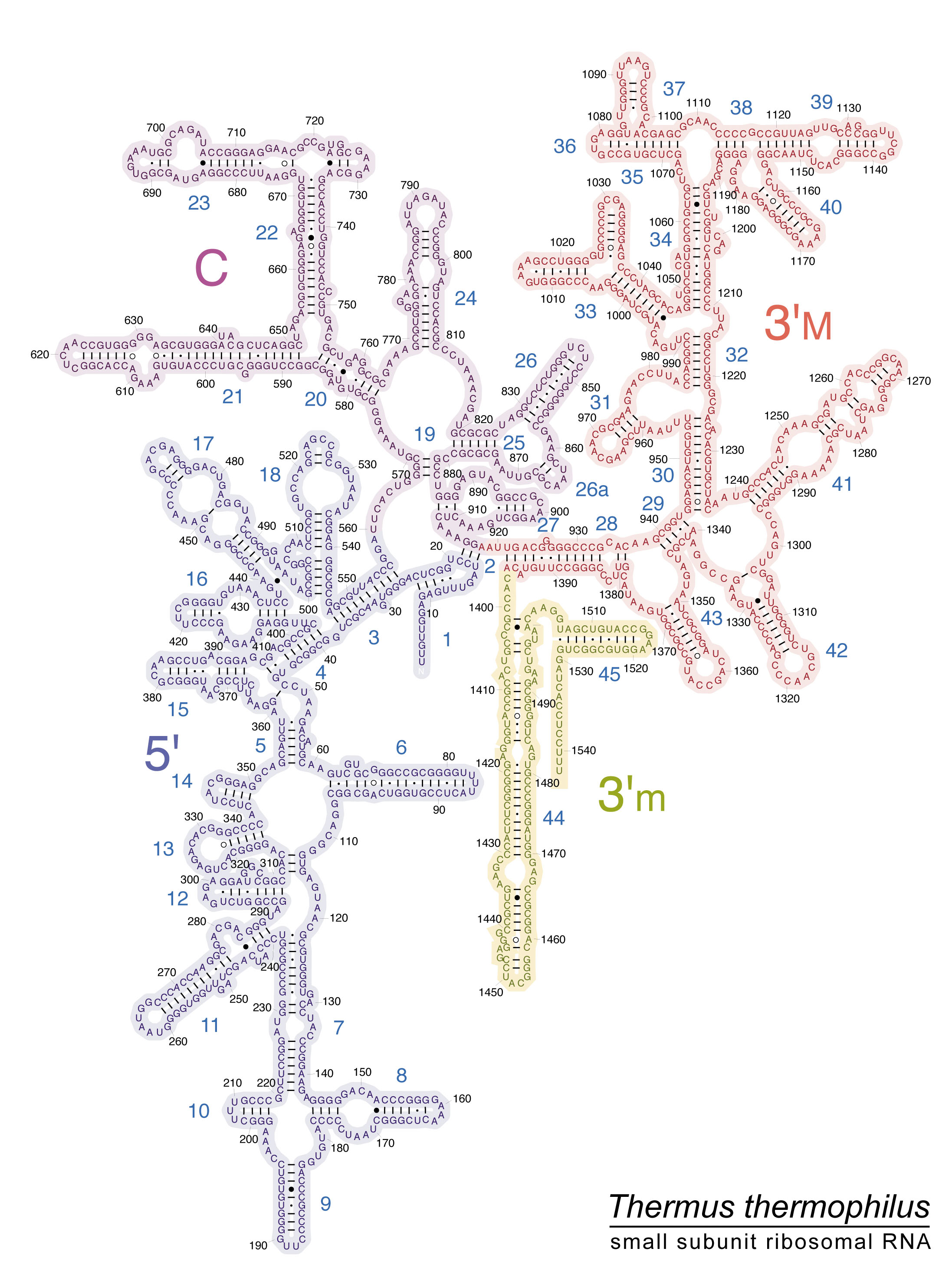
Thermus thermophilus 16S rRNA Secondary Structure (E. coli numbering)
JPG | TIFF | PDF
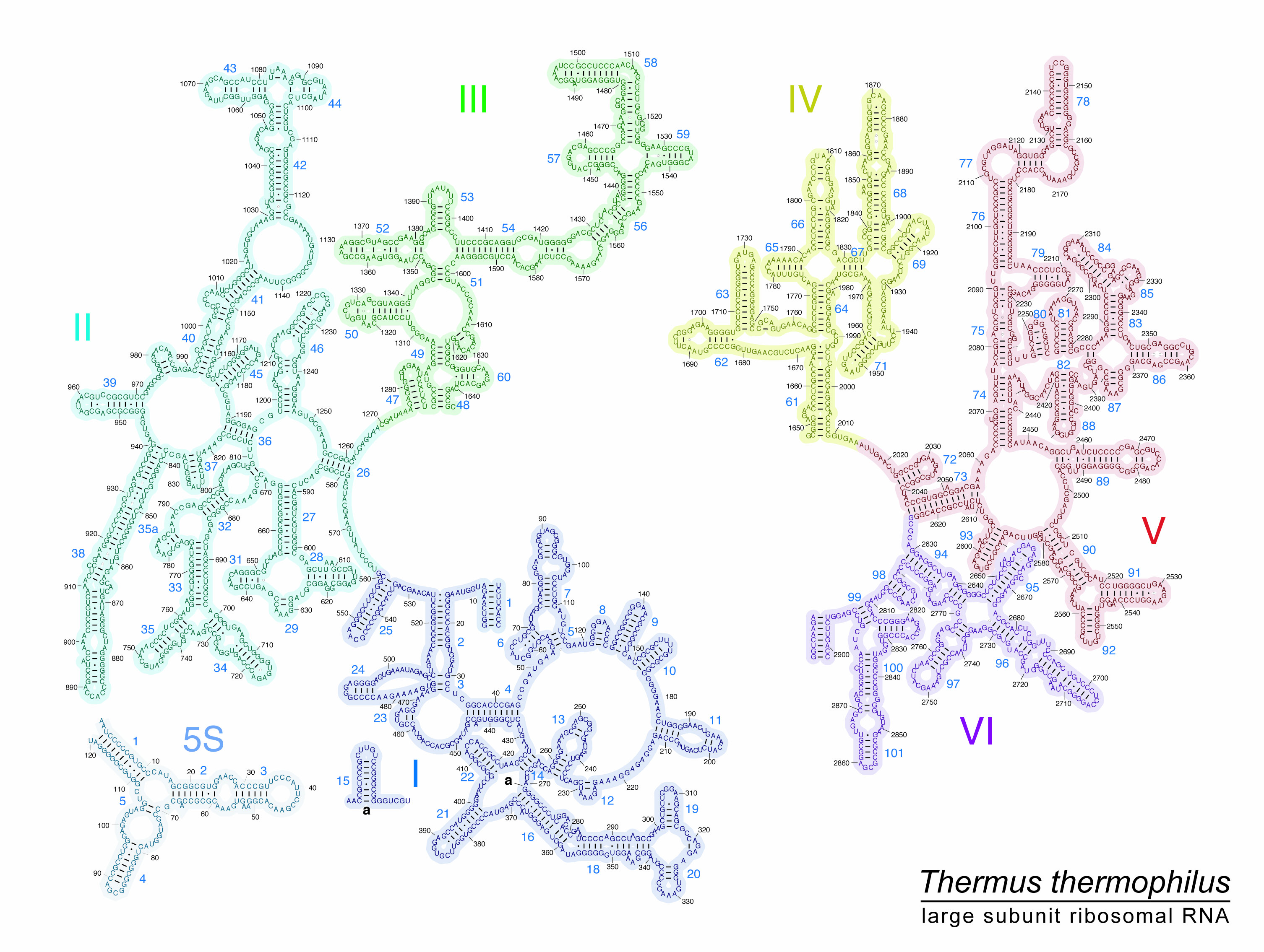
Thermus thermophilus 23S rRNA Secondary Structure (E. coli numbering)
JPG | TIFF | PDF
Escherichia coli 16S rRNA Secondary Structure
JPG | TIFF | PDF
Escherichia coli 23S rRNA Secondary Structure
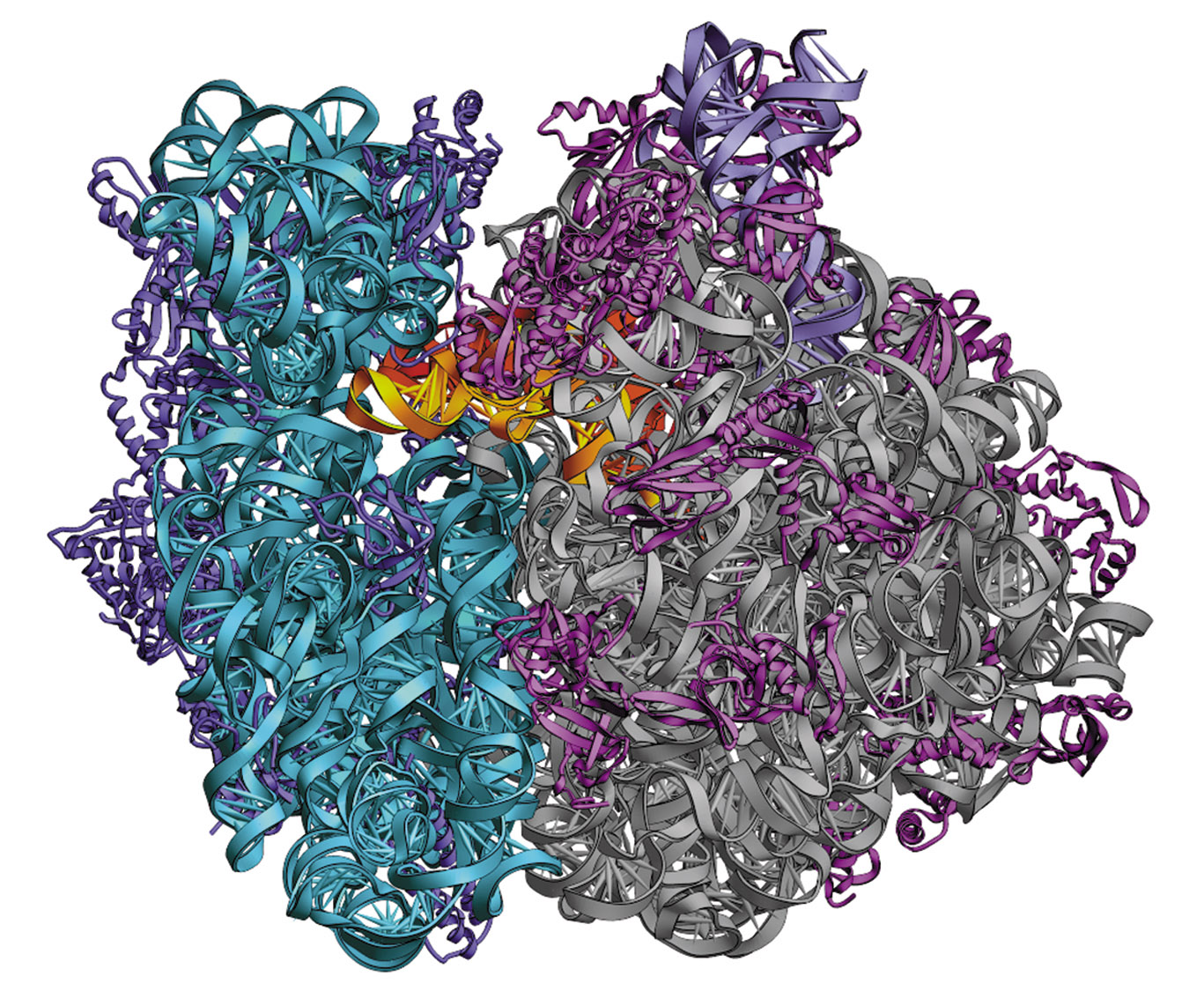
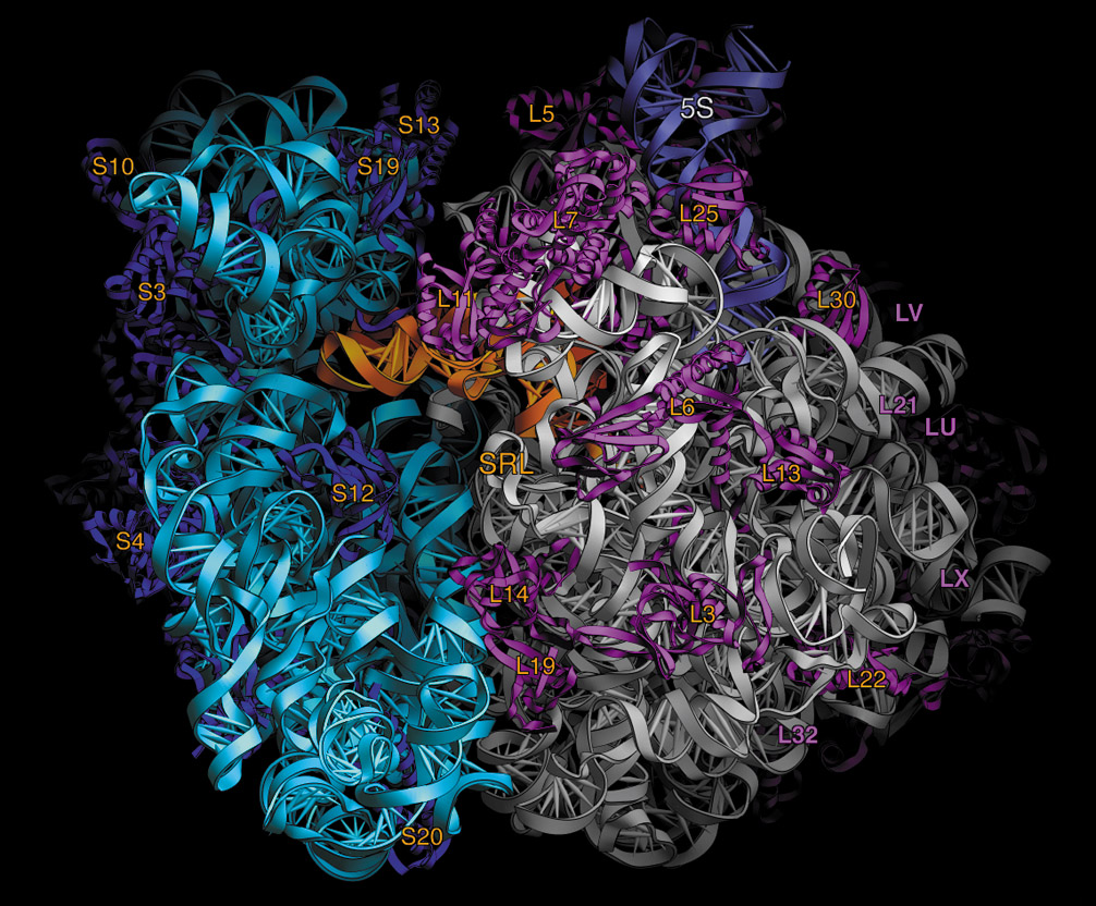
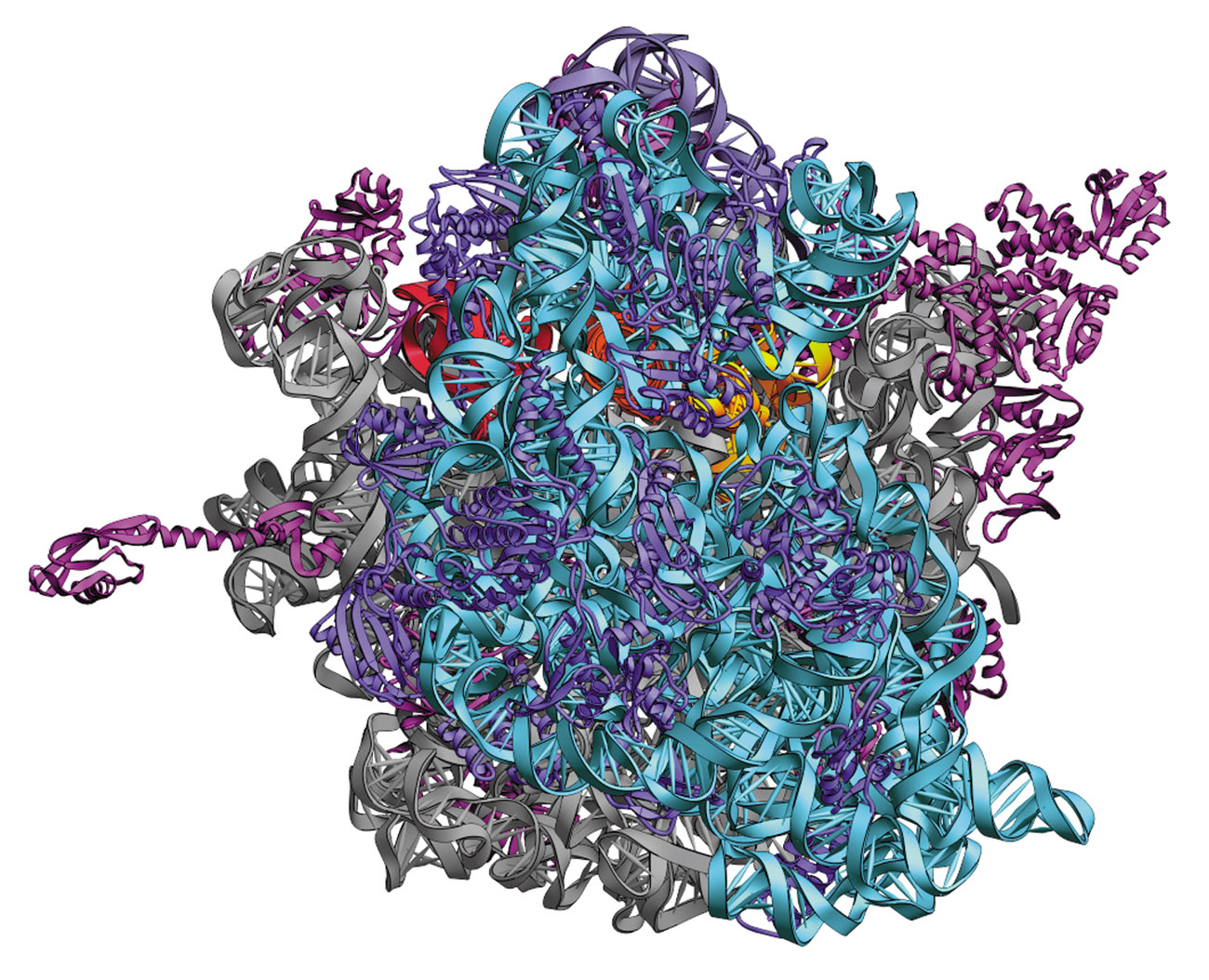
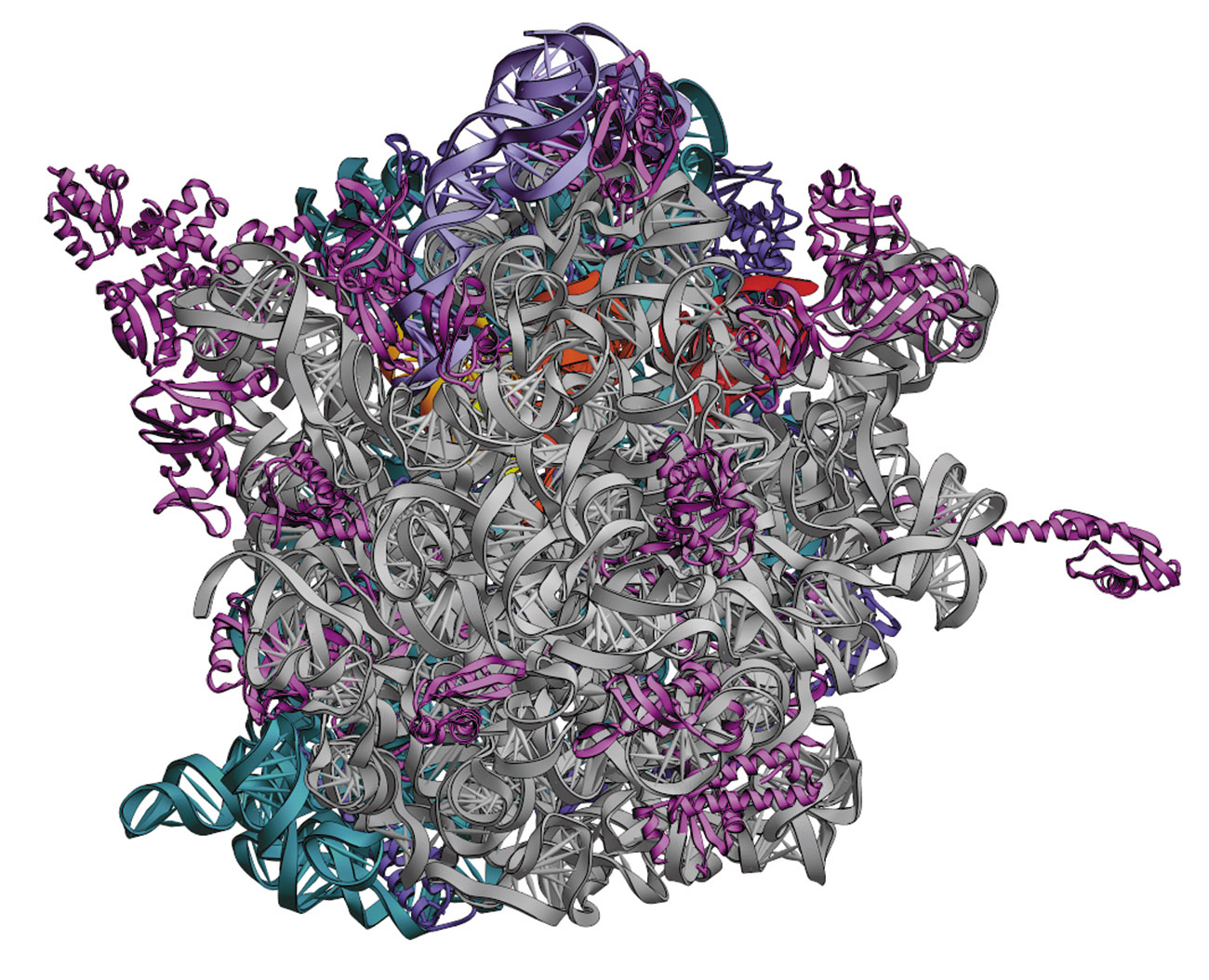
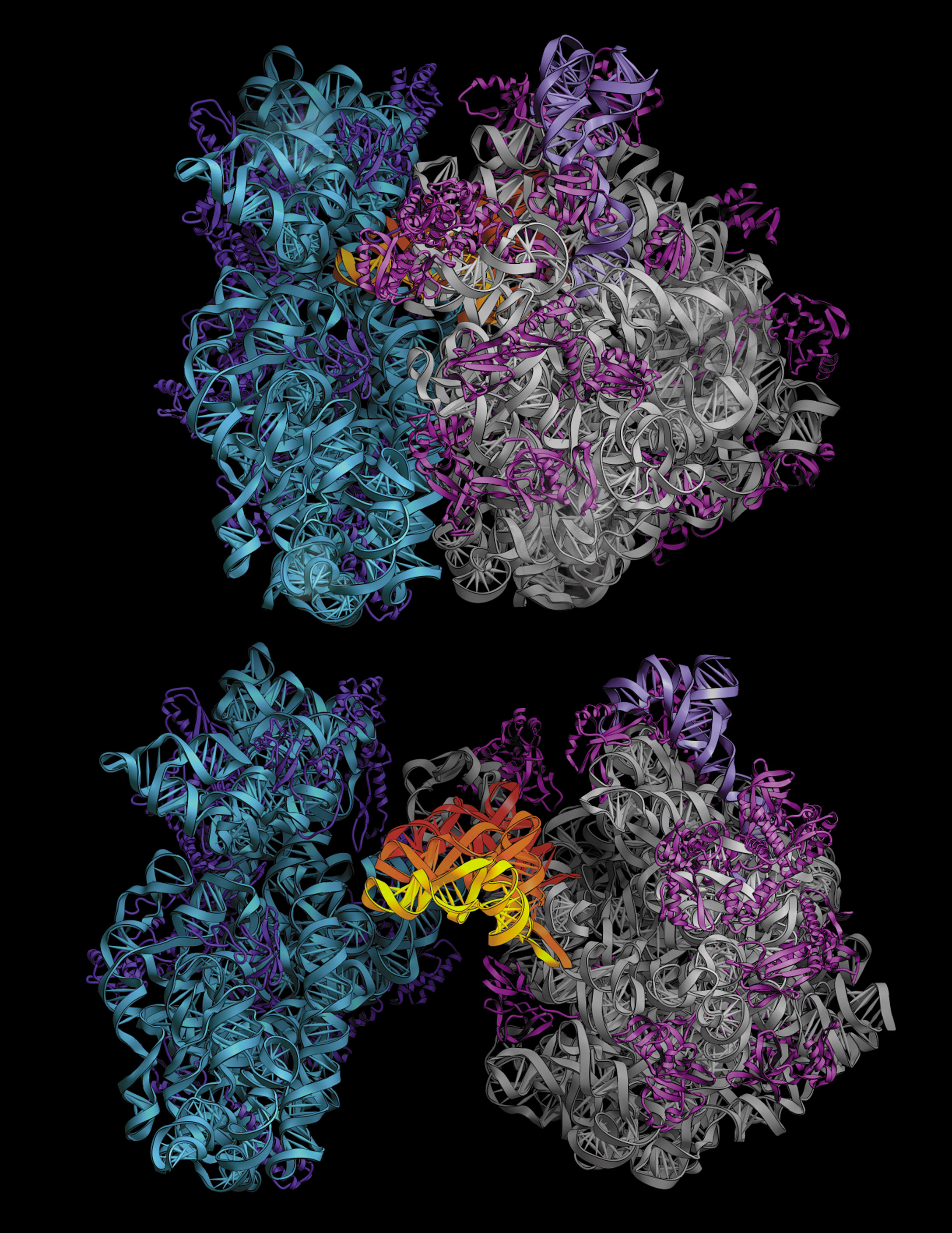
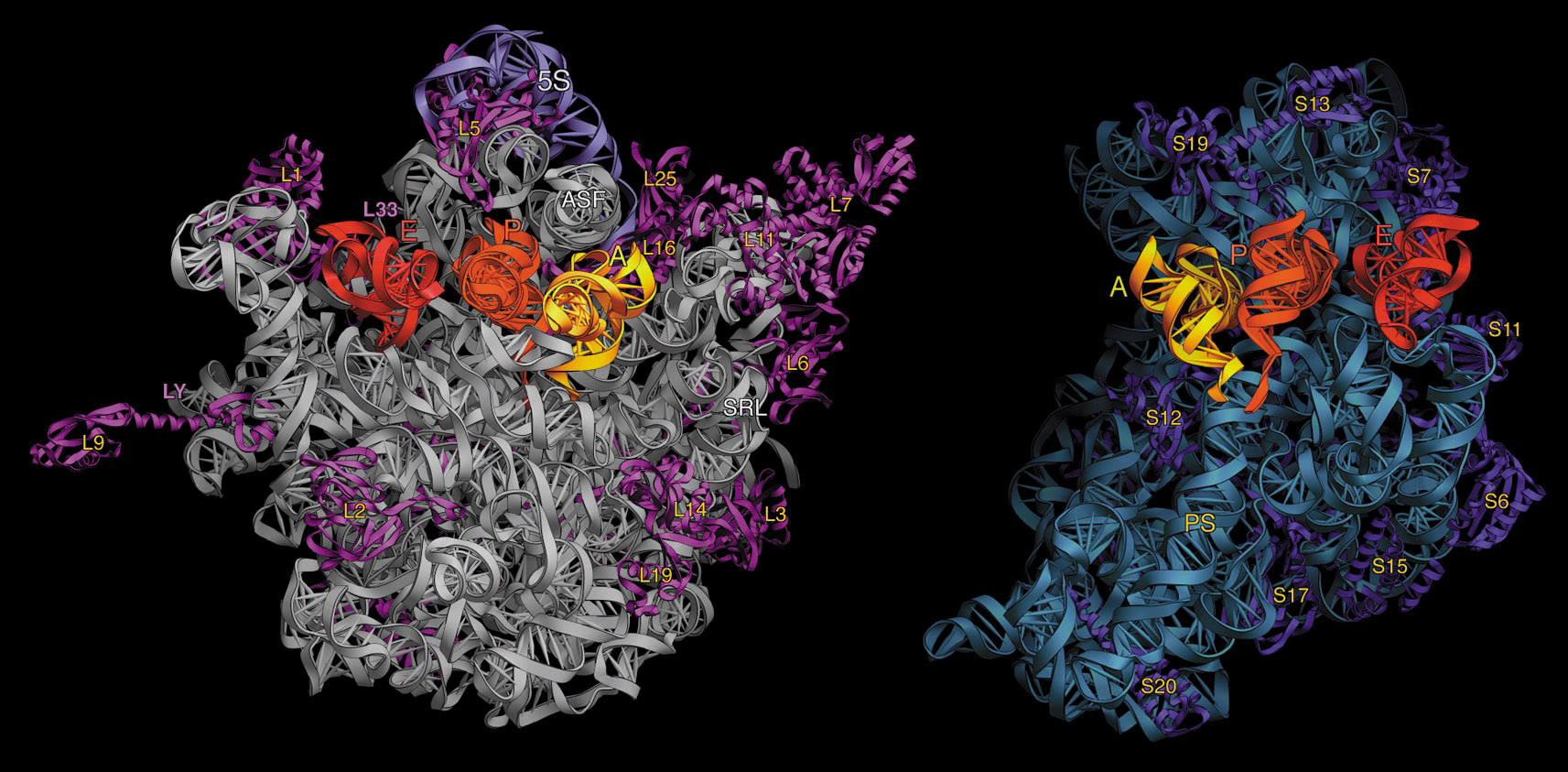
Ribosome Tertiary Structure
JPG | TIFF
Interface views of the 50S (left) and 30S (right) ribosomal subunits. with labels
Publication Covers
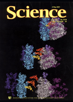
2001 Science Cover
2001 Cell Cover
1999 Science Cover
2002 FEBS Letters Cover
